Stemcelldoc's Weblog

April 21, 2014

ITB: Iliotibial Band Dysfunction and Treatment Options

At the Centeno-Schultz Clinic we approach pain and dysfunction in a systematic fashion which is outlined in Ortho 2.0.

We employ the acronym SANS:  symmetry, articulation, neurologic and stability.

This approach can be utilized for any joint.

The iliotibial band (ITB) is a fibrous band that extends from the hip (ilium) to the knee (tibia).

It is a critical in the stability of the hip and pelvis.

There are both anterior and posterior forces on the ITB.

Posteriorly it connects with the gluteus medius  muscle which when contracted pulls the ITB posteriorly.

Anteriorly the ITB connects with the tensor fasciae latae muscle which when contracted pulls the ITB anteriorly.

TFL_ITBand

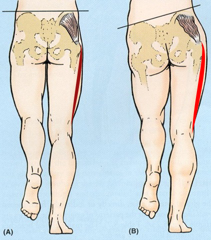
Biomechanics of ITB:  Supports and stabilizes the hip and pelvis when the opposite foot is suspended and off the floor such as when we are walking.

Lumbar spinal nerves provide essential information to the gluteus medius, tensor fasciae latae and other muscles that support the ITB.

Irritation or compression of lumbar nerves can result in a reduction in the nerve signal to the muscles and ensuing weakness.

This weakness can be witnessed on physical examination when the patient is asked to stand on one leg and the pelvis shifts.  Weakness in the ITB compromises the stability of the pelvis and hip with resultant shift of the pelvis as illustrated below.


ITB weakness


An excellent analysis and summary of the iliotibial band is presented below by Dr. Centeno.


April 19, 2012

Lateral Knee Pain: Iliotibial Band Syndrome

At the Centeno-Schultz Clinic we acknowledge that there are many causes of lateral knee pain.  An important case report was discussed in a previous blogOrtho 2.0 outlines the four key concepts: stabilization, articulation, neurological functions and alignment.

The iliotibial band (ITB) is a longitudinal fibrous reinforcement of one of the lateral leg muscles, the fascia lata.  It  originates from the anterior superior iliac spine region and extends down the lateral portion of the thigh to the knee.

Proximally it inserts into the lateral epicondyle of the femur and then passes to insert distally on the lateral aspect of tibia tubercle (gerdy’s tubercle).

When the knee is extended, the ITB is anterior to the lateral femoral condyle. When the knee is flexed the ITB is posterior to the lateral femoral condyle.

Actions of the iliotibial band include thigh flexion at the hip, abduction, medial rotation and  lateral stabilization the knee.

Iliotibial Band syndrome is an overuse phenomenon which is common in runners.  Typically pain occurs over lateral femoral epicondyle, which is caused by irritation and inflammation of the distal iliotibial band as it rubs against the lateral femoral condyle.

Differential diagnosis includes lateral meniscus tear,  popliteus,tendinitis, and patellofemoral pain syndrome.

The iliotibial band is a standard part of the diagnostic knee ultrasound conducted at the Centeno-Schultz Clinic.  In the ultrasound image below a normal  iliotibial band is identified by the white arrows.