Stemcelldoc's Weblog

May 30, 2012

Plantar Fasciitis: New Treatment Options

At the Centeno-Schultz Clinic we acknowledge that heel pain can be disabling.

Plantar fasciitis is the most common cause of heel pain and is caused by the  inflammation  of the plantar fascia at its insertion on the medial process of the calcaneus ( heel bone).  The plantar fascia is the thick connective tissue which supports the arch on the bottom of the foot.  It runs from heel bone (calcaneus) forward to the heads of the metatarsal bones.  Repetitive microtrauma of the plantar fascia due to alterations in foot bio mechanics is thought to be major pathology.

Intense sharp heel pain with the first couple of steps in the morning is a common presentation.  Other diagnostic considerations include:

  • Achilles tendon injuries

Lumbar radiculopathy

  • Calcaneal stress fracture
  • Calcaneal Bursitis
  • Contusions
  • Tarsal Tunnel Syndrome

In-0ffice MSK ultrasound at the Centeno-Schultz Clinic allows for radiation free method of visualizing the plantar fascia and diagnosing plantar fasciitis.

Treatment options for those who have not responded to rest and conservative therapy include IMS, ultrasound guided prolotherapy, PRP and percutaneous needle tenotomy. The latter is a non surgical procedure where under ultrasound guidance a small needle is placed into the fascia and small holes are created. A recent study by Vohra demonstrated greater than 80% improvement in 41 patients with chronic plantar fasciitis who underwent percutaneous needle tenotomy.  The ultrasound image below shows a needle directed into the plantar fascia in a patient with severe plantar fasciitis.

May 20, 2012

Anterior Hip Pain: Illiopsoas Tendinitis

At the Centeno-Schultz Clinic we acknowledge that hip pain can be disabling.

It may arise from osteoarthritis, avascular necrosis of the femoral head and femoral acetabular impingement syndrome.  Stem cell therapy is an alternative to traditional hip surgery.

Iliopsoas tendonitis can also cause hip pain which typically is anterior in location.  Tendinitis is inflammation of the tendon:  the rope-like tissue that connects muscle to bone.  Tendinitis is typically associated with an acute injury whereas tendinosis is chronic and associated with degeneration of the tendon.

Major causes include acute trauma and overuse from repetitive hip flexion.

The psoas and iliacus muscles originate from the lumbar spine and pelvis.  The muscles converge to form the iliopsoas muscle and insert onto the lesser trochanter.  The iliopsoas muscle functions as a hip flexor and external rotator of the femur.

Patients commonly present with anterior hip or groin pain.

Presentation may include shorten stride and anterior pelvic tilt.

Tenderness at the musculotendinous junction and insertion on tendon on the lesser trochanter is common.

The snapping hip sign or extension test is suggestive of iliopsoas injury.

At the Centeno-Schultz Clinic in-office ultrasound is an important diagnostic tool.  The noninvasive test typically demonstrates thickening of the tendon which is illustrated below.

Treatment options include prolotherapy, IMS, platelet derived growth factors and percutaneous needle tenotomy.

May 13, 2012

Carpal Tunnel Syndrome: Overview

At the Centeno-Schultz Clinic we acknowledge that wrist pain can be painful and limiting.

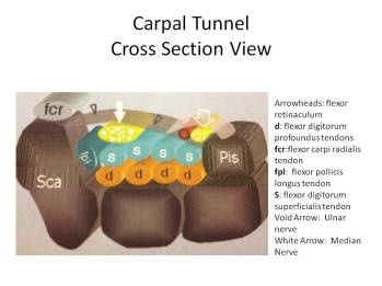
Carpal Tunnel syndrome (CTS) is an entrapment of the median nerve in the carpal tunnel causing pain and numbness.  The carpal tunnel is an anatomical compartment located at the base of the palm.  Nine flexor tendons and the median nerve pass through the carpal tunnel that is surrounded on three sides by the carpal bones that form an arch as illustrated below.

A successful case report utilizing  hydrodissection, a non surgical treatment has been discussed in a prior blog.

Patients commonly experience numbness, tingling, or burning sensations in the thumb, index, long and radial half of the ring finger.

Common clinical examinations include Phalen’s maneuver and Tinel’s sign.

Most cases of CTS are of unknown causes.

Diagnosis is often made by EMG/NCS study whereby needles are placed into muscles and electrical activity is evaluated.  The needles and the procedure  can be painful.  At the Centeno-Schultz Clinic MSK ultrasound is a non painful alternative.  A median nerve greater than 10mm in area indicates carpal tunnel syndrome.  A transverse ultrasound image of the median nerve is illustrated below.

Lateral Hip Pain: Beyond Trochanteric Bursitis

At the Centeno-Schultz Clinic we understand that hip pain can be debilitating.  Hip pain can arise from osteoarthritis, avascular necrosis of the femoral head(AVN),  labral tears and fractures.  Stem cell treatments are an alternative to traditional hip surgery.

Trochanterteric bursitis is an inflammation of the bursa on the outside aspect of the hip bone (greater trochanter).  The bursa, a fluid filled sac lies between the insertion of the gluteus medius and gluteus minimus  muscles into the  greater trochanter.

Other causes of lateral hip pain include inflammation(tendinitis) and degeneration(tendonosis) of the tendons.  MSK ultrasound is an office based evaulation at the Centeno-Schultz Clinic that allows for accurate diagnosis and treatment.  Trochanteric bursitis can be distinguied by diagnostic ultrasound so that appropriate therapy can be understaken.  Therapies include prolotherapy, PRP and Regenexx platelet derived therapies.

Below is a illustration of the 4 surfaces on the greater trochanter and the insertions of the gluteus medius and mimimus tendons.

May 6, 2012

Meniscus Tears: Common Types

At the Centeno-Schultz Clinic we acknowledge that a meniscus tear can not only be painful but can also impact your game.  Non surgical treatment options are outlined in this video.

The clinical success of stem cell therapy in treating a torn meniscus has been discussed in prior blog.

Meniscus are semilunar shaped cartilage wedges that act as shock absorbers between the thigh (femur) and tibia (shin) bones.  Menisci are triangular shape in cross section. Each covers approximately two-thirds of the corresponding articular surface of the tibia.  There is a medial (inner) and (lateral) meniscus in each knee-joint.

Meniscus tears are noted by how they look and where the tear occurs.

Common tears include longitudinal, bucket handle, flap, transverse and torn horn which are illustrated below.

May 5, 2012

Posterior Calf Pain: Medial Gastrocnemius Injury

At the Centeno-Schultz Clinic we acknowledge that there are many sources of posterior calf pain.  Meniscus injuries, osteochondral defects and ACL tears have been previously discussed as a cause of knee and posterior calf pain.  Stem cell therapy is an alternative to traditional knee surgery.

The gastrocnemius muscle is a large muscle in the posterior calf and is commonly injured in sport activities such as hill running, jumping and tennis.  The condition is sometimes referred to as tennis leg and is more common in men than woman.  Mechanism of injury is typically  an acute, forceful push-off with the foot.  An audible pop is often heard and is accompanied by pain in the calf with radiations into the ankle and restriction in range of motion.

The gastrocnemius muscle has two heads which originate respectively from the medial and lateral condyle.  The  two heads join and then merge with the soleus muscle-tendon complex to form the Achilles tendon.

The principle actions of the gastrocnemius muscle are ankle plantar flexion and knee flexion.

At the Centeno-Schultz Clinic MSK ultrasound is available within the office for evaluation of injuries.  Below is an ultrasound image of the posterior knee.