Stemcelldoc's Weblog

September 14, 2010

An NSAID a Day May Accelerate Knee Cartilage Degeneration

Non steroidal anti-inflammatory drugs (NSAID) are widely prescribed for joint pain.  What is the long-term effect of these common medications on knee cartilage?

Ding et al examined 395 patients with a mean age of 62 over 2.9 years in an effort to answer the question.  MRI’s were performed which evaluated both knee cartilage volume and cartilage defects.   Patients taking NSAID’s had increased knee cartilage defects and more loss in cartilage volume than patients who did not take NSAID.  Bottom line is that NSAID’s have a detrimental  affect on knee cartilage.

The Centeno-Schultz Clinic recognized this several years ago when mesenchymal stem cells in patients on NSAID’s failed to grow in a similar fashion as those who were not on these types of medication.  Mesenchymal stem cells, plasma rich platelets (PRP), prolotherapy and APC are several of the treatment options at the Centeno-Schultz Clinic.

July 27, 2010

Recovery from Stem Cell Therapy

Knee surgery often requires significant post procedure rehabilitation.

Microfracture, a common surgical procedure designed to create new joint cartilage, requires 6-8 weeks of non weight bearing after surgery.  This can lead to muscle weakness, joint stiffness and significant post procedure physical therapy.

CB, a professional athlete, despite undergoing two knee surgeries continued to have significant knee pain and limited range of motion.  CB was unable to climb stairs or run without significant pain.  Instead of undergoing an additional surgery, CB elected to use his own expanded stem cells using the Regenexx procedure.

 CB underwent additional therapies at the Centeno-Schultz Clinic which  included  prolotherapy to tighten loose ligaments and lumbar epidural injections with platelet derived growth factors. Compared to many orthopedic surgeries which require limited activity and extensive physical therapy, CB’s postprocedure restrictions were minor:  no high impact activity such as running and the use of an un-loader brace for 2 weeks. 

Several weeks following stem cell therapy, CB reports 85% improvement in his range of motion and knee pain.  He wanted to challenge himself so he tackled Vail Mountain.  Looking sharp…….

January 17, 2010

Is Knee Arthroscopy Effective ?

Knee arthroscopy is a common orthopedic procedure.  It is estimated that 1 million arthroscopic knee surgeries are performed annually in this country.

Knee arthroscopy involves making a small incision into the knee-joint through which a small camera and surgical tools are inserted.

Various parts of the knee are visualized which include the meniscus, the cartilage and supporting ligaments.  When damage is noted the area can be debrided:  rough surfaces are made smooth.

This is a significant advancement from the open surgeries performed in the 1980’s.

Is knee arthroscopy effective?

A recent article in the New England Journal of Medicine demonstrated that knee arthroscopy for osteoarthritis  provided no additional benefit to optimized physical and medical therapy.

A non surgical alternative to treat common orthopedic conditions is the use of your own mesenchymal  stem cells. The  Regenexx procedure allows patients to use their own mesenchymal stem cells to treat many knee, hip and shoulder conditions.

August 12, 2009

Enthesopathy: Fire in the Joint

fireTendons are fibrous connective tissue that attach a muscle to bone.

Ligaments attach bone to bone.

Inflammation of tendons and ligaments at their point of attachment onto bone are referred to as enthesopathies.  These can be very painful and lead to changes in gait and posture.

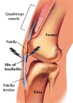
Common examples include golfer’s elbow and jumper’s knee.Pat_Tendonitis

Therapy is directed at reducing the increased tension in the affected muscle and decreasing the inflammation at the site of attachment of the tendon.  Unfortunately, all too often steroids are injected which may provide temporary relief but can weaken the ligament.  Repeat injections can lead to rupture.

At the Centeno-Schultz Clinic, enthesopathies are treated prolotherapy to strengthen the ligament.  Many patients who undergo stem cell therapy have enthesopathies which have contributed to their orthopedic conditions.  A complete and thorough evaluation and examination is our standard.

regenexx.mesenchymalstemcell.stemcelltherapy.kneecartilagetherapy.Centeno-Schultz Clinic.

July 30, 2009

Placement of Cells is Everything

Placement is Everything

Placement is Everything

Currently there are  numerous ways in which stem cells are being administered.

When injected intravenously a majority of the stem cells are trapped in the lungs.

When treating cartilage defects, does it matter how the cells are injected?

Koga et al.,clearly demonstrated that it does matter.  He  injected cells in a variety of ways including into the joint vs directly into the cartilage defect.  Cells placed directly into the cartilage defect resulted in more than 60% of the stem cells adhering to the area of damage and significant cartilage growth at 24 weeks.

A Colorado practice utilizes intermittent x-ray to inject autologous mesenchymal stem cells directly into areas of cartilage damage.  This technique avoids the risks and time associated with traditional surgical approaches. MRI images before and after stem cell therapy tell the story.

July 20, 2009

BMI and Road Wear

BMI not BMW.BMW

BMI is statistical measurement which compares a person’s weight and height.  It can be a useful tool to estimate a healthy body weight.

A recent study examined the knees of 336 overweight patients over 30 months.  20.2 % of the patients showed a slow loss of knee cartilage and 5.8% had rapid cartilage loss.  The Boston University researchers concluded that for every one-unit increase in body mass index, the chances of rapid knee cartilage loss increased 11%. Bottom line: stay lean.

If you have knee pain knee as a result of cartilage loss, there are non-surgical options.  Mesenchymal stem cell therapy involves the use of a patients own stem cells, avoids all of the risks associated with srugery and anesthesia and does not require extensive rehabilitation commonly associated with surgical procedures.

March 26, 2009

Synvisc Shoulder Injection Pain

Synvisc, also called hyaluric acid, is injectable medication used in the treatment of osteoarthritis.

Hyaluric acid is an important component of articulat cartilage.  It binds water and is responsible for the resilience of cartilage. Osteoarthritis, unfortunately erodes the articular cartilage of a joint giving rise to pain and reduction in range of motion.knee-cartilage-24

Synvisc when injected into a joint is commonly referred to as viscosupplementation.  It is thought that Synvisc supplements the viscosity of the joint fluid thereby lubricating the joint whereby in theory reducing the level of pain.

It can be injected in the knee, hip and shoulder joint.

The manner in which it is injected is critical.  Most clinician inject the joint without  x-ray guidance which can result in the deposition of the Symvisc outside of the intended joint.  This can and often times does create a significant amount of pain and affords the patient no therapeutic response since the medication was not delivered into the intended joint.  Jackson demonstrated that experienced orthopedics physicians are only successfully in entering the knee joint space 71% of the time when using an anterolateral approach WITHOUT x-ray.  At the Centeno-Schultz Clinic x-ray is always utilized with viscosupplementation to ensure accurate placement of the medication and to avoid the pain associated with injection of hyaluric  acid outside of the joint space.

If Snyvisc therapy fails to provide significant pain relief, patients now have the option of utilizing their own meseenchymal stem cells to regenerate cartilage in the knee, hip and shoulder.  It is a novel, needle-in, needle-out procedure with allows patients to avoid the risk of disease transmission as well as the trauma and rehabilitation associated with surgery.

Please review knee MRI below of patient who underwent the use of her own mesenchymal stem cells to regenerate cartilage.    Pre-treatment images are on the top and demonstrate a cartilage depth ranging between 0.2- 0.3cm.  Five months post therapy, the cartiage has increased on average by 1mm. 

Regeneration of knee cartilage through stem cell therapy

Regeneration of knee cartilage through stem cell therapy

Clinically the patient has had a significant reduction in her pain and an increase in her range of motion.  We just received an e-mail from her along with a picture of her daily activity:)

Back to Loving Life

Back to Loving Life

synvicinectionpain.synaviscinjectionpain.muscleatrophyinoneshoulder.physcialtherapyaftershouldersurgery.

March 10, 2009

Trendelenburg Test

trendelenburg-test1

Positive Trendelenburg Test with Weakness of Left Gluteal Muscles

The Trendelenburg test is a simple maneuver to evaluate the strength of the gluteus medius and gluteus minimus muscle.  It is performed by having the patient stand unassisted on one leg while the other leg is raised off the ground.  The examiner places their fingers on the posterior ilaic spine.  If there is a significant drop of the hip on the side of the raised leg,  their is weakness in the gluteus medius and minimus muscle.gluteal-muscles

The gluteus medius and mimus muscle are responsible for rotating the hip inward and extending it outward(abduction). 

They are critical in stabilizing the trunk and  keeping it in an upright position when one’s foot is raised while walking.

If there is weakness in the glueus medius and minimus there is structural instability with disproportionate forces placed on the hip and knee.  These forces can ultimately result in excessive wear of the joint cartilage and pain. knee-cartilage-22Often times surgery is the recommended therapy.

At the Centeno-Schultz Clinic we emphasis structural integrity.  If a patient has a positive Trendelenburg, they will be scheduled for therapy to restore gluteal strength which  typically is in the form of intramuscular stimulation(IMS).  If there has been hip or cartilage damage, a novel technique using your own stem cells can used to assist in the regeneration the cartilage.

March 5, 2009

New Knee Cartilage

The knee is covered with cartilage.

Normal and Damaged Knee Cartilage

Normal and Damaged Knee Cartilage

Cartilage is living material that produces glycosaminoglycans (GAG’s),  which allow for lubrication as well as holding onto water within the cartilage.

mogul-skiing

Bye, Bye Cartilage Cells

Maintenance of cartilage is a dynamic process.  Each time we cycle, ski the bumps or run, a number of cartilage cells perish.  The cells are then replaced by circulating mesenchymal stem cells which differentiate into new cartilage cells.  When the amount of damage exceeds repair, the cartilage starts to dry up and degenerate.  When the balance is reversed so that repair exceeds the damage, the cartilage remains healthy and full of GAG’s.  It is the balance that is key.  Unfortunately, all the issues regarding regeneration and degeneration of cartilage have yet to be fully understood.  What is known is that with age, the number of circulating repair cells (mesenchymal stem cells) decreases.

At Regenexx we attempt to restore a normal balance by injecting  autologous bone-marrow-derived mesenchymal stem cells into the area of damaged cartilage.  This is performed under x-ray guidance and a small needle.  Regenexx is unique in that we utilize a patient’s own stem cells, and therefore avoid the risk of disease transmission or rejection.  Unlike bone marrow aspirate concentrate therapies, we expand the number of mesenchymal stem cells so as to afford the patient the best outcome.

The MRI below outlines one of our recent knee successes.  This is a patient who had undergone microfracture surgery on her knee due to significant loss of cartilage, without success.  The top images are prior to the procedure,  bottom images are 16 months after therapy.  The red arrows identify breaks in the cartilage (white area in the gray cartilage).  The yellow arrows point to the areas of previous cartilage damage which have now been repaired.  Note the white areas which indicated cartilage damage have significantly resolved.  Clinically, the patient is now pain free and was last seen somewhere in Vail.

Knee Cartilage Before and After Regenexx Procedure

Knee Cartilage Before and After Regenexx Procedure

February 10, 2009

Valgus and Varus Alignment

Valgus and varus describe the alignment between two anatomical segments.  Valgus  refers to outward angulation of the distal segment of a bone or joint.  Varus is the opposite of valgus and refers to inward angulation of the distal segment of a bone or joint. To better understand these concepts, see the  illustration below.   A line is drawn along the long axis of the proximal segment.  Another line is drawn along the long axis the distal segment.  When comparing the two lines,  focus on the the distal segment’s alignment with respect to the proximal segement. valgus2

 The illustration on the left depicits valgus in which the distal segment of the joint is angulated outward.

The illustration on the right is an example of varus.varus-alignment

What is the significance?    Look at the x-rays below.valgus-x-ray1


Misalignment of one bone in relation to other places disproportionate forces on the joint with resultant excessive wear and tear on the meniscus, ligament and cartilage.  At the Centeno-Schultz Clinic we emphasis correct joint aligment through various therapies including IMS, prolotherapy, physical therapy and the use of unloader braces.  If there has been excessive wear of the cartilage,  there is a novel technique which allows patients to use there own stem cells.


Valgus;  – femur internally rotated, and tibia externally rotated.  Problems:  maliagnilgment as results from asymmetric wear of the posterior aspect of the lateral femoral condyle  patellofemroal tracking problems. patellar maltracking. posterolateral instability

Older Posts »