Stemcelldoc's Weblog

March 30, 2014

Lateral Arm Numbness and Weakness: Consider Axillary Nerve Dysfunction

At the Centeno-Schultz Clinic we acknowledge that shoulder pain can compromise quality of life and athletic endeavors.

Our online book, Ortho 2.0 highlights a systemic evaluation of joint pain and dysfunction.  The approach is termed SANS.

The N is SANS denotes nerve dysfunction.

Axillary nerve dysfunction can be a cause of lateral arm numbness and shoulder weakness.

The Axillary nerve arises from the posterior cord of the brachial plexus, C5 and C6.

Course:  It crosses the antero-inferior aspect of the subscapularis muscle, passes behind the arm through the quadrilateral space, winding around the neck of the humerus ending in two major trunks:  anterior and posterior trunk.

Axillary nerve

Innervation:  Teres Minor and Deltoid

Sensory: Lateral deltoid also known as the regimental badge area.

Axillary-Nerve

Injury can occur at several sites along the nerve path:

Origin of the posterior cord

Anterior-inferior aspect of the subscapularis muscle

Quadrilateral Space:

suprascapular_and_axillary_nerves_02

The quadrilateral space is bounded superiorly by the teres minor muscle, inferiorly by the teres major muscle, medially by the long head of the triceps, and laterally by the humeral shaft.

Common presentation is dull intermittent ache or pain that is localized in the posterior and lateral shoulder.  Symptoms are exacerbated by active and resisted abduction and external rotation of the humerus.

Quadrilateral space syndrome is an affliction of middle-aged men and it presents classically in the throwing athlete and those with a history rotator cuff surgery.

Ultrasound examination is can be significant for atrophy of the teres minor muscle which is illustrated below.

Ultrasound Image of Teres Minor in Quadrilateral Space

 injury at several sitesgin from the posterior cordAnteroinferior aspect of the subscapularis muscle and shoulder capsuleQuadrilateral space

March 26, 2014

Common Causes of Chronic Knee Pain: A systematic Approach

At the Centen0-Schultz Clinic we utilize a comprehensive approach to patient care.    Our online ORTHO 2.0 discussess a systematic approach to common orthopedic conditions.

Don’t assume that loss of cartilage is the source of all knee pain.

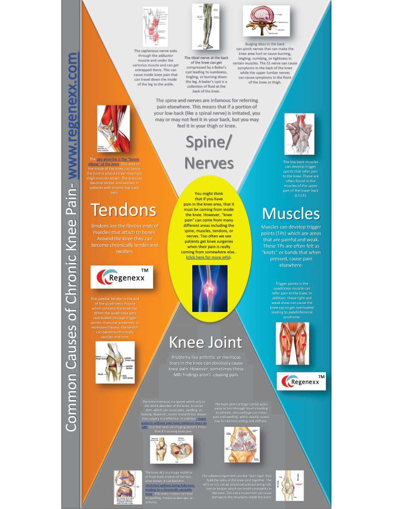
Below is an infographic which outlines the common causes of knee pain.

Tendons:  patellar, distal quadriceps and pes anserine.

Spine:  Lumbar disc disease with compression of L5 and or S1 nerve root.

Peripheral nerves:  compression and or irritation of saphenous and tibial nerves.

Muscles:  Dysfunctional muscles resulting in weakness or imbalance.

Ligaments:  key stabilizers of the knee joint include the ACL, MCL and LCL.

Knee Joint:   medial and lateral meniscus

Bottom Line:  prior to undergoing any treatment ensure that the principal pain generator has been identified to maximize clinical results.


Common-causes-of-Chronic-knee-pain-Infographic

March 25, 2014

ACL Tear Stem Cell Study: Free Care for Those Who Meet Criteria and can Travel

tree with clouds

The Centeno-Schultz Clinic is a research based medical practice that acknowledges the importance of research.

Our publications to date are multiple and easily identified in the U.S. Library of Medicine.

Stem cell therapy is a successful alternative to traditional surgery in the treatment of ACL tears.

Successful cases of ACL repairs have been discussed in prior blogs with comparisons of pre and post MRI’s illustrating the healing afforded by autologous stem cell treatment.

Regenexx currently has an ACL stem cell study which is available for patients that qualify and can travel to our Broomfield clinic.

Full Inclusion criteria is listed on web site and includes:

  1. Physical examination consistent with lax ACL ligament (Anterior Drawer Test)
  2. Abnormal Telos Arthrometer measurement
  3.  Positive diagnostic MR imaging of the affected knee with at least 1/3 of the ACL ligament at any area along its length having high signal on MRI PDFS/Fat Sat images.

Exclusion criteria is also listed and includes:

  1.  A massive ACL tear or one that includes more than 2/3’rds of the ligament that’s retracted.
  2.  Previous surgery to the affected ACL
  3. Concomitant meniscus tear or cartilage injury that occurred at the same time as the as the ACL tear and which is considered a pain generator.

If you or family or friends are interested please contact us at 303 963-9528 or complete the Regenexx candidacy form.


March 17, 2014

Regenexx ACL Procedure Featured in Outside Magazine

outside magazine logo

Regenexx ACL procedure is dynamic new technology that enables a patient to use their own stem cells to heal ACL tears.  This procedure was featured in Outside Magazine.

Extreme skier and founder of DPS Ski, Stephan Drake, tore his ACL while skiing a steep face in Alaska.  He rejected surgical repair and opted for Regenexx ACL at the Centeno-Schultz Clinic in Colorado.

Regenexx ACL is a alternative to traditional knee surgery.  Bone marrow derived stem cells are injected under direct visualization into the ACL.  Patients are fitted with knee brace and undergo 4 weeks of physical therapy.

Complications of ACL surgeries have been discussed previously and include graft failure, infection and acceleration of arthritis.

Successful clinical cases with detailed pre and post treatment MRI’s outlining improvement have been presented in previous blogs.

It is a new day in orthopedic medicine with new opportunities.  Not long ago open heart surgery with bypass and coronary grafting was the standard of practice.  Then interventional cardiology with a new skill set including balloon angioplasty became the new standard of care making coronary bypass surgery a less prevalent and less attractive procedure.

March 12, 2014

Supercharged Platelet Rich Plasma

The Centeno-Schultz Clinic utilizes a special advanced platelet rich plasma (PRP).

The injection of platelet rich plasma is a successful treatment option for joint, tendon, ligament, muscle and joint injuries.  The concentrated blood platelets contains critical growth factors that increase the body’s ability to repair itself.

In most clinics and hospitals and clinics, PRP is created by a bedside centrifuge that separates the plasma and concentrates the platelets.  Bedside centrifuges typically create a PRP with platelet counts of 3-5 times the normal.  Unfortunately they also contain white and red  blood cells which can cause inflammation.

bedside

Are there differences in PRP treatments?

YES!

Regenexx has a superior PRP  called Regenexx SCP (Super Concentrated Platelets).  It is created by cell biologists in a state of the art laboratory and results in a 10-20 fold concentration of platelets.  Higher concentration of platelets has been shown to have a positive effect on the stem cells responsible for repairing damaged tissue.

White and red blood cells within platelet rich plasma can result in excessive inflammation, pain, restriction in range of motion and inhibition of  repair cells.

Bedside centrifuges are not adept at removing white and red blood cells whereas cell biologists at Regenexx are very capable of separating these cells. The image below demonstrates the cardinal difference.  The PRP on the left is red in color due to the red blood cells whereas the Regenexx SCP on the right is amber in color due to the successful removal of most red and white blood cells.

scp_vs_prp

PRP is an effective treatment of many ligament, tendon, joint and muscle injuries.  There are differences in platelet rich plasma concentrates.  Regenexx SCP which is produced in a state of the art lab by a cell biologists affords a much higher level of platelets with reduced red and white blood cell contaminates which ultimately results in  best possible outcomes.

March 9, 2014

Another Reason to Avoid NSAID: Delayed healing in rotator cuff after surgical repair

burning-flames-yellow-fire1

At the Centeno-Schultz Clinic we acknowledge the negative impact of NSAID on repair.

NSAID have been reported to affect bone metabolism,  fracture healing and increase the risk of sudden death heart attack.

Unfortunately NSAID medications continue to be prescribed for musculoskeletal pain including pain subsequent to surgical rotator cuff repair.

Does this medication make a difference in healing?

Checkik in a recent article examined the impact of NSAID on rotator cuff healing after surgical repair.  39 rates underwent rotator cuff injury and surgical repair.  Group A received meloxicam for the first 10 days whereas Group B recived meloxicam from postoperative day eleven.  Group C received saline injections.

Results:  Group B displayed lower maximal load which is consistent with delayed healing.

Bottom Line:  Avoid NSAID as they delay healing.

March 3, 2014

Groin Pain: Consider Iliopsoas Tendinitis

At the Centeno-Schultz Clinic we acknowledge the pain and restriction in motion associated with groin pain.

Inflammation of the iliopsoas tendon can be a cause of groin and anterior hip pain.  Treatment options include prolotherapy and platelet rich plasma injections.

The psoas and iliacus muscle originate from the lumbar spine, converges to form the iliopsoas muscle and its tendon  attaches onto the lesser trochanter of the femur.

Iliopsoas Tendon

The musculotendinous junction can be found in a groove between the anterior inferior iliac spine and the iliopenctineal eminence.

Major causes of iliopsoas tendinitis are  trauma and overuse resulting from repetitive hip flexion.

In adolescents the combination of inflexible hip flexors and tightness of the iliopsoas muscle and tendon can lead to anterior pelvic tilt and increased stress on the lower lumbar discs and facets.

Groin or anterior thigh pain is a common presentation as illustrated below in red.

iliopsoas referral pain pattern

Physical examination may be significant for anterior pelvic tilt, tenderness of the iliopsoas tendon and a snapping hip sign.

At the Centeno-Schultz Clinic  a diagnostic ultrasound is utilized to confirm the diagnosis.  Below is an ultrasound image identifying the iliopsoas tendon and adjacent femoral artery.

Iliopsoas Tendon Ultrasound Image

March 2, 2014

Successful Repair of Anterior Cruciate Ligament Tear with Stem Cells

At the Centeno-Schultz Clinic we recognize that tears of the Anterior Cruciate Ligament (ACL) can be game changers.  Surgical repair is associated with significant complications which include infection, graft failure and alteration of biomechanics.  Many patients who undergo surgical repair do not return to high level competition.

Stem cell therapy is a viable alternative to traditional surgical repair.   The Regenecxx  SD procedure enables a patient to forgo surgery, the lengthy rehabilitation and utilize their own stem cells to heal tears in the ACL.

HK is a case in point.  He is a 44 y/o athletic weekend warrior who sustained a hyper-extension injury with near complete tear of the ACL.  He declined surgical repair and underwent Regenexx SD.  6 months post injection of his stem cells he had complete return to normal activities and sports.  His professional and scholastic endeavors delayed follow-up MRI which accounts for the gap in pre and post imaging.

Pre and post stem cell treatments are posted below.  The pre-injection images are on the left and the post injection images are on the right.  The first images are taken from the side (sagittal) whereas the second set are frontal (coronal).

The thigh bone is on top (femur) and the tibia is below.  The ACL stretches across the knee-joint and prevents excessive forward motion.  The ACL is outlined in dashed yellow lines.  On the pre-injection images note the disorganized quality of the ACL fibers and the disruption of the ACL towards the top.  On the post injection images the ACL fibers are well-organized, tight and uniform extending from the tibia to the femur.  The formal MRI report post stem cell injection notes no tear in the ACL which is consistent with HK’s clinical results.

Keel Successful ACL Stem Sag

Keel Successful ACL COR

In 2014 stem cell treatment is an alternative to traditional surgery for tears of the ACL.