Stemcelldoc's Weblog

January 6, 2012

Quadriceps Tendon Pain

The Centeno-Schultz Clinic offers PRP, prolotherapy and bone marrow and platelet derived stem cell therapies.  An understanding to function and stability is essential and is covered in Ortho 2.0.  Direct visualization using MSK ultrasound or x-ray is our standard to ensure accurate placement and to avoid the complications associated with blind knee injections.

Muscle and tendon function is critical to the knee joint health. A brief review of the anterior compartment is helpful.

The quadriceps is a group of large muscles in the front of the thigh.  It consists of four major muscles:
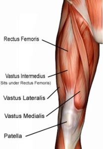
Rectus femoris: A large muscle that covers most of the other, deeper quadriceps muscles.

Vastus lateralis

Vastus intermedius

Vastus medius

A tendon is a fibrous band of connective tissue that connects the muscle to the bone.  The quadriceps tendon attaches the quadriceps muscles to the patella.

Quadriceps control knee extension and stabilize the kneecap (patella).

Pain can arise from the quadriceps tendon due to inflammation, chronic degeneration or tear.  A tear can be either partial or complete and is usually the result of trauma.  Tendon weakness predisposes to tears.  Conditions that can lead to tendon weakness include  quadriceps tendonitis, chronic diseases, steroid use, immobilization and the use of a class of antibiotics called fluoroquinolones.

July 22, 2011

Treatment of Lateral Elbow Pain with PRP

Pain on the outside aspect of the elbow is referred to as lateral epicondylitis or tennis elbow.  It can be quite disabling affecting both the quality of life and range of motion.

Lateral epicondylitis is caused by irritation of the common extensor tendons as they insert onto the bony surface of the elbow.

PRP (platelet rich plasma) has become increasing popular as non surgical treatment in common orthopedic conditions.

Has it been used in the treatment of elbow pain?

Yes!

Peerbooms demonstrated PRP to be more effective than steroids in the treatment of lateral elbow pain.  100 patients were randomly assigned treatment with either PRP or steroids.  At one year 73% of patients treated with PRP reported significant improvement vs 49% in the steroid group.

Steroid use has been associated with significant side effects including ligament and cartilage compromise and cell death (apoptosis).

At the Centeno-Schultz Clinic advanced cell treatments including PRP are used in the treatment of lateral elbow pain and other common orthopedic conditions including meniscus tears, supraspinatus tears,  osteoarthritis of hip, knee and anklesMSK ultrasound and or x-ray is used in injections to ensure accurate placement of platelets.

October 22, 2009

PRP and Athletic Injuries

 Wanted to share the treatment of a local sports personality who responded well to PRP therapy.

Patient suffered an injury to the semimembranous which is one of the three muscles that comprise the hamstring.  He had significant pain in the back of this thigh which limited his ability to run.  he had point tenderness on examination with swelling and bruising.

 The semimembranosus muscle allows us to flex the knee, extend the hip and rotate our hip inwardly when the knee is flexed.

semimembranosus

MRI demonstrated a tear in the muscle which is identified by the white arrow.

MRI of semimembranous

Patient underwent  a concentrated injection of his own platelets into the area of injury to stimulate repair.  This concentrate is referred to as platelet rich plasma (PRP).  Platelets are rich in growth factors which have been demonstrated to promote natural healing. After two injections the patient had 80% improvement and was able to return to playing.  PRP therapy affords patients a natural healing treatment with minimal downtime and the potential for significant results. 

PRP has been successfully used in professional athletes as witnessed by the treatments of Hines Ward  prior to the Superbowl.

PRP is just one of the regenerative therapies utilized at he Centeno-Schultz Clinic.

October 15, 2009

Steroids: Patients Beware

Filed under: Cervical Spine, Lumbar Spine — Tags: , , , , — stemcelldoc @ 2:51 pm

Steroids are commonly used to treat pain.  Common examples include cortisone injections into knees, elbows, hips and lumbar spine.

There is increasing concern by many authors about the side-effects of steroid injections.

Hossain demonstrated that steroids were capable of shutting down normal repair and maintenance functions in the joint.(Apoptosis) 

Nakazawa demonstrated that steroids can turn off the production of catilage.

Wong identified a correlation between steroid use and bone death. (avascular necrosis of the femoral head)

At the Centeno-Schultz Clinic we too are concerned and have changed our treatment protocols from large dose steroid injections to low dose.  Most steroid injections use 6 milligrams of steroid.  At Centeno-Schultz Clinic we use a very dilute concentration of steroid which is compounded for our clinic.  A nanogram is 1/1000 th of a milligram.  Steroid injections at Centeno-Schultz Clinic  are between 50 to 100 nanograms.  This dilute solution has positive effects on joint health by increasing the concentration of growth factors and stimulating stem cell production of cartilage.

Other options for the treatment of pain include prolotherapy, PRP and stem cell therapy.

February 26, 2009

The Importance of Blood Supply

Blood supply to a given structure provides essential nutrients for growth, maintenance and repair.  Some structures such as the heart have a rich blood supply with multiple arteries.  Other structures have a very limited, tenuous blood supply which places them at risk for impaired growth, repair and potential cellular death. The lumbar intervertebral disc is such a structure.  While it is the cornerstone of our spine and bears the weight of our bodies as we walk, its blood supply is extremely limited.

What does this mean?  If injuries occur the lumbar disc has limited capability of repairing itself due to its very limited blood supply.  The result is a slow insidious degeneration of the lumbar disc characterized by reduction in disc height and signal.  The MRI  below on the left illustrates a degenerative L5/S1 disc.  The blue arrow identifies the spinal cord, the red arrow is pointing to the cerebral spinal fluid and the black arrow identifies the L3/4 disc.  Note the L3/4 disc has a white signal within the disc which represents hydration.  It also is identical in height and brightness to the disc above it.  Both of these discs are normal.  The white arrow identifies a degenerative L5/S1 disc which is black in color with reduction in height in comparison to the adjacent discs.

Degeneration of lumbar spine

Degeneration of lumbar spine

 With injury there is also a propensity to develop bulges since the integrity of the side wall (annulus fibrosis) is compromised. 

Disc Bulge

Disc Bulge

 If sufficient damage occurs, a disc bulge can progresses to a disc herniation where an portion of  the inner contents of the disc (nucleus pulposus) are extruded.

Lumbar Disc Herniation

Lumbar Disc Herniation

In a previous blog I discussed the importance of platelets and the four major growth factors they contain:   Platelet-derived growth factor (PDGF), transforming growth factor beta (TGF-b), vascular endothelial growth factor (VEGF) and epithelial growth factor (EGF). Vascular endothelial factor is responsible for angiogenesis (the creation of  new blood vessels).  These new blood vessels can provide essential nutrients to repair damaged tissue.  At Regenexx we injected concentrated VEGF adjacent a degenerative disc with the hope of improving blood flow and initiating repair.  Please see MRI results below.  On the left are MRI images of the L5/S1 disc prior to therapy.  Note there is a reduction in height and brightness of the L5/S1 disc.  On the right are MRI images of the same L5/S1 disc after therapy.  Note an increase in the height and the signal of the disc.  There is significant improvement in the disc height and signal.  What is the significance?  This is a patient who despite prior back surgery continued to have pain.  After therapy at Regenexx the patient had near complete resolution of pain as well as MRI evidence of lumbar disc repair.

Before and after therapy of L5/S1 Disc

stemcelltherapy.lumbardegenerativediscdisease.alternativestolumbarsurgery.CentenoSchultClinic.VEGF

February 24, 2009

Platelet Rich Plasma

Filed under: Stem Cell Basics — Tags: , , — stemcelldoc @ 4:57 pm

What do Pittsburgh Steelers’ Hines Ward and Troy Polamalu have in common?  They have both used their own blood to treat ligament and tendon injuries.  The therapy is called platelet-rich plasma (PRP) and involves the injection of concentrated platelets and plasma to the area of damage.  Instead of injecting steroids, which can lead to tissue damage and tendon rupture, physicians are now injecting a patient’s own blood products to stimulate healing.

What is PRP?  It is a concentrate of a patient’s own platelets.  In our bodies, normal platelet counts range between 150,000/μl and 350,000/μl with an average of  200,000/μl.  Animal and human studies have demonstrated that soft tissue healing enhancement only occurs when the concentration of platelets (PRP) is greater than 1,000,000/μl.  This is a concentrate of 5x the amount normally present in our bodies.

How is it made?  A small amount of a patient’s own blood is placed in a centrifuge which separates the red blood cells from the platelets.  A teaspoon of the remaining substance is injected into the damaged tissue.

How does it work?   Platelets contain several significant growth factors, which enhance tissue repair.  The most important growth factors in PRP are platelet-derived growth factor (PDGF), transforming growth factor beta (TGF-b), vascular endothelial growth factor (VEGF) and epithelial growth factor (EGF).

What are the advantages?  It is a non-surgical therapy utilizing a patient’s own cells, and therefore is not associated with allergic reaction or transmission of disease.  At the Centeno-Schultz Clinic we use PRP for repair of ligament, tendon and muscular injury.

February 4, 2009

Lateral Epicondylitis

Lateral epicondylitis otherwise known as tennis elbow is a overuse injury involving the extensor muscles that originate on the bony prominence (epicondyle) on the outside (lateral) aspect of the elbow.  It is more properly termed a tendinosis that specifically involves the origin of the extensor carpi radialis brevis muscle.  Nirschl and Pettrone attributed the cause of lateral epicondylitis to be tearing in the origin of the extensor carpi radialis brevis (ECRB) muscle.

flexor-carpi-radicalis

The extensor carpi radialis brevis (ECRB) muscle originates from the lateral epicondyle. It functions to move the wrist so that the hand moves away from the palm and towards the thumb.

Many patients with tennis elbow are involved in work or recreational activities that require repetitive use of the forearm muscle. Pain can be  severe and burning  localized on the outside part of the elbow.   In most cases the pain starts slowly and escalates over weeks or months.

Conservative therapy includes limiting repetitive use of the forearm.  Orthotics can only help diminish symptoms of tennis elbow.lateral-epicondyle-brace1

These can reduce symptoms by resting the muscles and tendons.  The surgical procedure involves removing diseased tendon tissue and reattaching normal tendon tissue to bone.

lateral-epidcondyle-surgery

Alternative therapies include prolotherapy, platelet-rich plasma and stem cell therapy.  At Regenexx patients are able to use their own stem cells to regenerate torn ligaments and tendons.